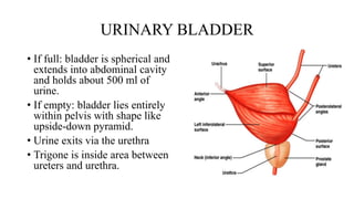
Urinary Bladder Earths Lab Biology Diagrams Urinary bladder (sagittal view) The urinary bladder is a pelvic organ that collects and holds urine before urination. It serves as a temporary reservoir for urine produced by the kidneys.When empty, it lies completely within the pelvic cavity, but enlarges upward into the abdominal cavity when full.It is the most anterior pelvic organ, located just behind the pubic bones and pubic symphysis.
It is a small tube leading from the urinary bladder to the exterior of the body. In males, the urethra extends through the penis, is about 20 cm in length, and is divided into three regions: the prostatic, membranous and spongy urethra. In males, the urethra also functions to transport semen, formed by the testes and accessory sex glands. The urinary bladder and urethra are pelvic urinary organs whose respective functions are to store and expel urine outside of the body in the act of micturition (urination). As is the case with most of the pelvic viscera, there are differences between male and female anatomy of the urinary bladder and urethra. In our entire urinary system series, the urinary bladder and urethra represent the

Urinary bladder & urethra: Anatomy, location, function Biology Diagrams
The urethra in both males and females begins inferior and central to the two ureteral openings forming the three points of a triangular-shaped area at the base of the bladder called the trigone (Greek tri- = "triangle" and the root of the word "trigonometry").The urethra tracks posterior and inferior to the pubic symphysis (see Figure 4). Issues with the urethra are more common in people who have male urethral anatomy. Urinary tract infections, including catheter issues. Urinary tract infections are very common. This test lets your provider look into your urethra and bladder with a cystoscope (a small scope with a camera). X-rays and/or ultrasound. These imaging tests allow

The urethra is the vessel responsible for transporting urine from the bladder to an external opening in the perineum.. It is lined by stratified columnar epithelium, which is protected from the corrosive urine by mucus secreting glands.. In this article, we shall look at the anatomy of the male and female urethra - their anatomical course, neurovascular supply, and any clinical correlations.
Medicine LibreTexts Biology Diagrams
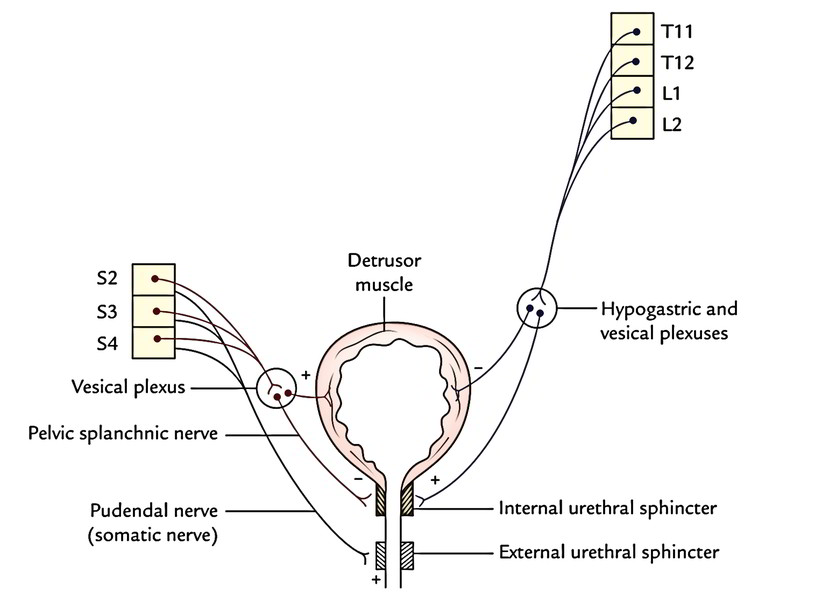
The nerves alert a person when it is time to urinate, or empty the bladder. Urethra. This tube allows urine to pass outside the body. The brain signals the bladder muscles to tighten, which squeezes urine out of the bladder. At the same time, the brain signals the sphincter muscles to relax to let urine exit the bladder through the urethra.